
2026 Top Immunohistochemistry Staining Types
Immunohistochemistry staining remains a practical bridge between tissue structure and molecular diagnosis. In 2026, laboratories are refining established methods while adopting more multiplex and image-assisted approaches. The strongest choices still depend on specimen quality, antibody validation, and clinical purpose. Newer does not always mean better.
This guide examines the top immunohistochemistry staining types used in research, pathology, and translational medicine. It covers chromogenic staining, immunofluorescence, multiplex panels, double staining, and automated workflows. Each method creates a different visual result. A brown DAB signal may highlight one protein clearly, while fluorescent channels can reveal several markers within the same cell. Small details matter, including fixation time, antigen retrieval, tissue thickness, and counterstain balance.
Experienced technicians know that staining is rarely perfect. Background signals can resemble weak expression. Uneven sections can mislead interpretation. Controls may pass, yet the clinical picture may remain unclear. That is why reliable practice combines validated protocols, positive and negative controls, documented quality checks, and specialist review. Digital pathology can improve consistency, but it cannot replace judgment.
This overview draws on established laboratory principles and current diagnostic practice. It also recognizes an important limitation: staining performance varies among tissues, platforms, antibodies, and laboratories. Results should therefore be interpreted within validated procedures and relevant clinical context. The goal is not to promote one universal technique. It is to clarify when each staining type may provide useful, defensible evidence.
Fundamentals of Immunohistochemistry Staining
2026 Top Immunohistochemistry Staining Types: Fundamentals of Immunohistochemistry Staining
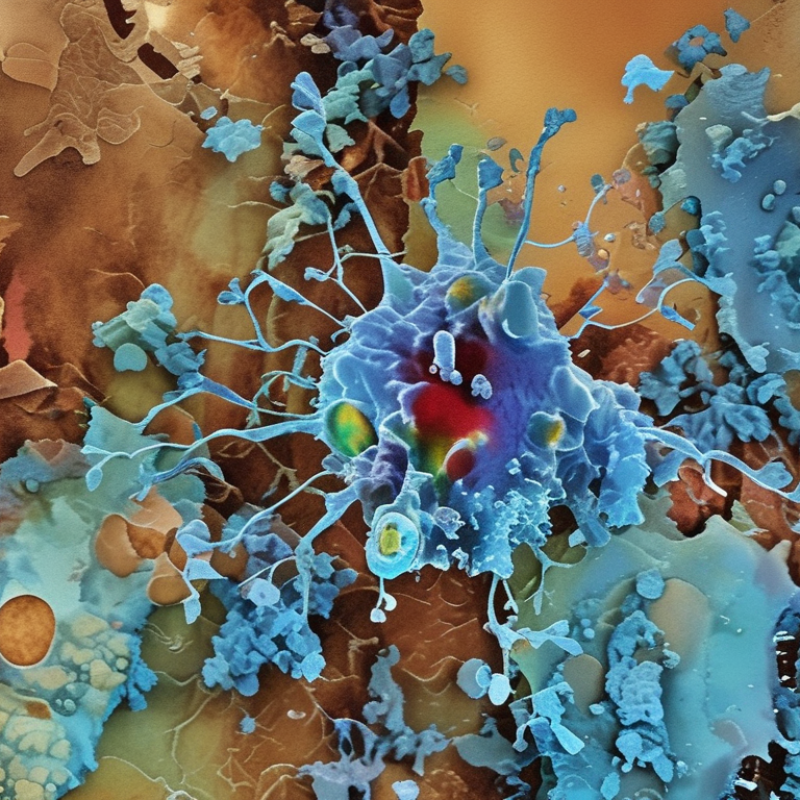
Immunohistochemistry, or IHC, links antibody binding with visible tissue signals. It helps pathologists locate proteins inside preserved cells. The main staining types include chromogenic, fluorescent, direct, indirect, and multiplex methods. Chromogenic IHC uses an enzyme reaction to create colored deposits, often brown or red, on a slide. Fluorescent IHC uses light-emitting labels and can show several targets. However, fluorescence may fade, and interpretation can become difficult.
The IARC Global Cancer Observatory reported about 20 million new cancer cases worldwide in 2022. That burden increases the need for consistent tissue-based diagnosis. Reliable IHC begins with fixation, section thickness, antigen retrieval, antibody selection, and control tissues. A weak signal may reflect poor retrieval, not absent protein. I have seen technically attractive slides mislead interpretation. That detail deserves more attention.
Tips: Keep positive and negative controls beside patient sections. Record fixation time and retrieval conditions. Use clear scoring rules before reviewing results. Multiplex staining can reveal protein patterns, but more markers do not always mean better evidence. The College of American Pathologists emphasizes validated procedures, documented controls, and staff competency in laboratory quality practices. Small deviations matter. A rushed protocol can produce clean-looking, unreliable findings.
2026 Top Immunohistochemistry Staining Types - Fundamentals of Immunohistochemistry Staining
A practical comparison of widely used immunohistochemistry and immunostaining formats
| Staining Type | Core Principle | Detection System | Typical Signal | Main Strengths | Important Limitations | Common Applications |
|---|---|---|---|---|---|---|
| Direct Immunohistochemistry | A labeled primary antibody binds directly to the target antigen in the tissue section. | Enzyme- or fluorophore-labeled primary antibody. | Colored precipitate or fluorescence at the antigen site. | Simple workflow, fewer incubation steps, and lower risk of secondary-antibody cross-reactivity. | Usually less sensitive because there is little or no signal amplification. | Abundant antigens, rapid research screening, and selected routine stains. |
| Indirect Immunohistochemistry | An unlabeled primary antibody binds the antigen, followed by a labeled secondary antibody directed against the primary antibody. | Enzyme- or fluorophore-labeled secondary antibody. | Chromogenic or fluorescent signal concentrated at target locations. | Greater sensitivity, flexible secondary-antibody selection, and signal amplification from multiple secondary antibodies. | Additional steps increase processing time and may produce background from nonspecific secondary binding. | Routine tissue profiling, diagnostic pathology, and research-based antigen localization. |
| Peroxidase–Anti-Peroxidase (PAP) | A primary antibody is followed by a secondary antibody and a soluble peroxidase–antiperoxidase immune complex. | Horseradish peroxidase-based immune complex. | Usually a brown reaction product after oxidation of a chromogenic substrate. | Historically important amplification method with good tissue localization. | More complex and generally less sensitive or less convenient than many modern polymer-based systems. | Classical immunohistochemistry protocols and selected archival methods. |
| Avidin–Biotin Complex (ABC) | Biotinylated secondary antibody binds a preformed avidin–biotin–enzyme complex, concentrating enzyme molecules at the target. | Biotin–avidin or biotin-binding protein complex with an enzyme label. | Commonly a brown, insoluble enzyme reaction product. | High sensitivity and strong signal amplification compared with direct staining. | Endogenous biotin in some tissues can cause background unless adequately blocked; multiple steps are required. | Research immunohistochemistry, tissue antigen mapping, and historical diagnostic protocols. |
| Polymer-Based Detection | A polymer backbone carrying multiple enzyme molecules is linked to the antibody-detection system, often without biotin. | Enzyme-labeled polymer coupled to a secondary antibody or antibody-binding reagent. | Usually a permanent colored precipitate, such as brown or red, depending on the chromogen. | High sensitivity, shorter workflows, and reduced endogenous-biotin-related background. | Very strong amplification can increase nonspecific staining if antibody concentration and blocking are not optimized. | Modern routine surgical pathology and high-throughput tissue evaluation. |
| Chromogenic Immunohistochemistry | An enzyme linked to the antibody system converts a soluble substrate into an insoluble colored product at the target site. | Most often peroxidase or alkaline phosphatase enzyme activity. | Brown, red, blue, or other visible precipitate that can be viewed by bright-field microscopy. | Permanent slides, familiar microscopic interpretation, and compatibility with routine histology. | Limited spectral multiplexing and possible interference from endogenous pigments or enzyme activity. | Clinical diagnosis, biomarker assessment, and long-term slide archiving. |
| Immunofluorescence | Antibodies carrying fluorescent labels identify antigens through emitted light after excitation at an appropriate wavelength. | Fluorophore-conjugated primary or secondary antibodies. | Bright fluorescent signal detected by fluorescence or confocal microscopy. | High spatial resolution, multicolor analysis, and suitability for colocalization studies. | Photobleaching, tissue autofluorescence, limited long-term signal stability, and need for specialized imaging. | Cell signaling, protein colocalization, immune-cell phenotyping, and research microscopy. |
| Multiplex Immunohistochemistry | Multiple antibodies detect several targets in the same tissue section using distinct colors, fluorophores, or sequential signal-development steps. | Sequential chromogenic detection, fluorescent labels, or signal-amplification chemistry. | Multiple spatially resolved signals that can be analyzed individually or together. | Preserves tissue context while measuring several biomarkers and cell populations in one section. | More demanding optimization, antibody cross-reactivity risks, spectral overlap, and complex image analysis. | Tumor microenvironment studies, immune profiling, spatial biology, and translational research. |
| Immunocytochemistry | The same antibody-based antigen-detection principles are applied to cultured cells, cell smears, or cytology preparations rather than tissue sections. | Direct or indirect enzyme-based and fluorescent antibody systems. | Chromogenic or fluorescent signal within individual cells. | Useful for cell morphology, protein localization, and controlled experimental systems. | Cell fixation and permeabilization can alter morphology or antigen accessibility; tissue architecture is limited. | Cultured-cell experiments, cytology specimens, and subcellular localization studies. |
Related Posts
-
How to Optimize Immunohistochemistry Staining for Best Results?
-
Understanding the Top Stem Cell Research Innovations and Their Impact?
-
What Are the Top 10 Minimal Residual Disease Tests?
-
2026 Best Recombinant Antibody Applications and Benefits?
-
What is the Role of Secondary Antibodies in Immunoassays?
-
Top 10 Innovations in 3D Cell Culture You Should Know About?