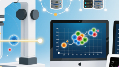
2026 Top Cell Imaging Systems for Global Buyers?
Choosing among the 2026 top cell imaging systems requires more than comparing resolution figures. Global buyers need dependable evidence, practical workflows, and support that survives daily laboratory pressure.
Eric Betzig, a Nobel Prize-winning pioneer in super-resolution microscopy, once said, “I think the most exciting thing about microscopy is that it allows you to see things that you couldn’t see before.” His words still guide purchasing decisions. A system should reveal meaningful biology, not merely produce attractive images.
This overview examines leading platforms for live-cell imaging, fluorescence microscopy, high-content screening, and advanced 3D analysis. It considers optical performance, acquisition speed, automation, environmental control, software usability, and long-term service. These details matter when cells must remain healthy inside a controlled chamber for hours, while hundreds of wells are recorded and analyzed.
The best system depends on the experiment.
A pharmaceutical screening laboratory may value robotic throughput and reproducible segmentation. A university core facility may need flexible objectives, multiple fluorophores, and simple user training. A clinical research group may prioritize traceability, data protection, and validated workflows.
Specifications can mislead. Higher magnification does not always produce better biological insight. Faster cameras may create unnecessary data. Artificial intelligence can reduce analysis time, but weak training data may introduce quiet errors.
This guide compares systems with those limitations in mind. It also recognizes an uncomfortable reality: no platform is perfect. Buyers may still face software learning curves, expensive upgrades, or uneven regional service. Careful testing with representative samples remains essential before purchase. The strongest choice is the system that produces reliable, interpretable results in the buyer’s actual laboratory, not the one with the longest feature list.
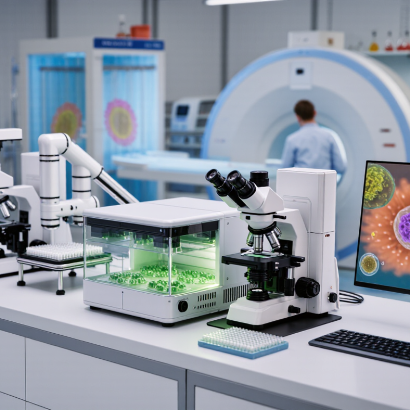
What Cell Imaging Systems Are and How They Work
Cell imaging systems combine illumination, optics, cameras, automation, and analysis software. They capture cellular changes that ordinary observation cannot measure reliably. Brightfield imaging records shape and density. Fluorescence imaging detects tagged structures, proteins, or viability signals. Some platforms also use phase contrast, time-lapse capture, and automated plate handling.
The workflow is practical. A sample sits in a dish or multiwell plate. Controlled light reaches the cells, while lenses focus the emitted or transmitted signal onto a camera. Software then corrects background, identifies objects, and measures features such as area, intensity, movement, and cell count. According to Grand View Research’s 2024 analysis, the global cell imaging market is expected to expand strongly through 2030, driven by drug discovery, disease research, and high-content screening. MarketsandMarkets also identifies automated imaging and image analysis as major growth areas in its 2024 market assessment.
Yet automation does not remove judgment. Poor focus, uneven illumination, cell stress, or unsuitable exposure can distort results. A beautiful image may still be scientifically weak. This matters for global buyers comparing systems in 2026. Check environmental controls, optical resolution, software validation, data export, service coverage, and local training. The best system is not always the most complex one. It should match the assay, sample volume, and operator skill. Even then, segmentation can fail on crowded cells. Human review remains necessary. That limitation deserves more attention.
How to Compare the Top Cell Imaging Systems in 2026
How to Compare the Top Cell Imaging Systems in 2026
Comparing cell imaging systems requires more than checking camera resolution. Start with your biological question, sample type, and daily workload. A system for fixed cells may perform poorly with fragile live cultures. Check optical resolution, illumination uniformity, autofocus speed, and environmental control. These details affect images captured at the edge of a 96-well plate. Ask for raw image files, not only processed examples. Reliable evaluation depends on evidence.
Throughput matters when experiments involve thousands of wells. Measure acquisition time, focusing accuracy, storage demands, and analysis speed. Review whether the software supports segmentation, tracking, and export to common data formats. Confirm how easily researchers can audit analysis settings. Service quality also matters. Request training records, maintenance schedules, validation procedures, and response times. A lower purchase price may hide expensive consumables, upgrades, or specialist labor. No system wins every application. Pilot testing can reveal uncomfortable assumptions.
Tips: Compare identical samples on each system. Use stained and unstained controls. Test low-contrast cells, crowded wells, and uneven illumination. Record hands-on time, failed runs, and data review effort. A short trial is useful, but it may not represent long-term reliability. Ask independent users about recurring problems. Keep a written scoring sheet, and let multiple operators repeat the test.
Leading Cell Imaging Systems for Global Research Buyers
2026 Top Cell Imaging Systems for Global Buyers
Leading cell imaging systems help research teams capture consistent data across laboratories, climates, and workflows. In practical evaluations, image quality matters, but repeatability matters more. Look for stable brightfield and fluorescence imaging, dependable autofocus, and clear measurements across 96-well plates. Environmental control can protect live-cell studies during long observations. Simple software also reduces training time for new operators.
Tips: Request raw-image exports, calibration records, and regional service details. Test the system with your own cells before purchase. Ask how updates affect existing analysis methods. A short trial often reveals more than a polished demonstration.
Global buyers should examine compatibility with microscopes, incubators, plate formats, and laboratory information systems. Strong systems support automated exposure, cell counting, confluence analysis, and secure data storage. They should also provide understandable reports for researchers and auditors. However, no platform fits every experiment. A system optimized for fixed samples may disappoint in long-term live-cell work. I have seen teams overvalue resolution while overlooking workflow delays, maintenance access, and staff training. That mistake can become expensive. Local regulations, import conditions, language support, and replacement-part availability deserve equal attention. Reliable performance is built through suitable configuration, documented methods, and honest validation rather than impressive specifications alone.
Global Purchasing, Compliance, and Integration Considerations
2026 Top Cell Imaging Systems for Global Buyers?
Global buyers should evaluate cell imaging systems beyond image quality. A strong platform must fit local electrical standards, laboratory workflows, and import requirements. Check voltage, connector types, software language, and service coverage before issuing a purchase order. These details seem minor until installation stops beside a packed loading dock.
Compliance also needs practical review. Ask for safety documents, calibration records, cybersecurity information, and clear data ownership terms. Confirm whether image files can be stored locally or transferred across borders. Procurement teams should verify customs classifications and required shipping documents with qualified local specialists. Rules differ by destination, and assumptions create expensive delays.
Tips: Build a site-readiness checklist before comparing prices. Include room temperature, bench space, network access, training, and maintenance response time. Request a sample workflow using your actual cell types and reporting format. No shortlist is perfect. In our experience, integration problems often appear after acceptance testing, not during demonstrations. A system may produce excellent images yet frustrate researchers through slow exports or complicated user permissions. Leave budget for validation, staff training, and unexpected software adjustments. Reliability is not only a specification; it is daily usability under real laboratory pressure.