2026 How to Choose the Best Live Cell Imaging Microscope for Your Research?

The field of live cell imaging has advanced significantly, enabling researchers to observe cellular processes in real time. The live cell imaging microscope plays a crucial role in this evolution. With an estimated market growth rate of 7.4% from 2021 to 2026, these microscopes are becoming indispensable in cell biology and related fields. The ability to see cells in their natural environments provides insights that fixed imaging techniques cannot offer.

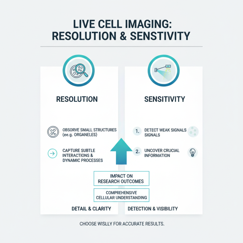
Choosing the best live cell imaging microscope can be challenging. Researchers must consider several factors, including resolution, sensitivity, and the types of fluorescent probes available. A recent survey indicated that over 65% of scientists prioritize image clarity when selecting their equipment. This aligns with findings from major reports, suggesting that optical performance directly impacts experimental outcomes. However, budget constraints and technology limitations often force scientists to compromise.

Every research project is unique, and so are its needs. There is no one-size-fits-all solution in this specialized area. Continuous reflection on experimental goals and technological advancements is essential for making informed decisions.

2026 How to Choose the Best Live Cell Imaging Microscope for Your Research?

Understanding the Fundamentals of Live Cell Imaging Technology

Live cell imaging technology has transformed biological research. This technique allows scientists to observe cells in real-time, providing insights into cellular processes. Understanding how live cell imaging works is essential for researchers. Microscopes harness advanced optics and illumination methods, enabling detailed visualization of live specimens.

Imaging quality depends on multiple factors. Resolution and contrast are crucial for clarity. Researchers often face challenges regarding light exposure. Excessive light can harm delicate cells, leading to inaccurate results. Balancing illumination and cell health requires careful consideration. Environmental controls, like temperature and humidity, also play a role. Maintaining optimal conditions is vital for realistic observations.

Choosing the right microscope can be overwhelming. Each model offers different features, such as camera quality and software capabilities. It's important to assess your specific needs. Many users often overlook aspects like ease of use and support. Exploring these elements helps in making informed decisions. As technology evolves, staying updated is necessary. Adapting to new advancements can enhance research outcomes significantly.

2026 How to Choose the Best Live Cell Imaging Microscope for Your Research?

Feature Importance Recommendations
Objective Lens High resolution and light-gathering capability Choose lenses with numerical aperture > 1.3
Illumination System Ability to minimize phototoxicity Consider LED or laser light sources
Imaging Speed Critical for dynamic processes Select systems with high frame rates
Live Cell Compatibility Essential for maintaining cell health Look for temperature and CO2 control options
Software Features Ease of use and analysis capabilities Use software with robust image analysis tools

Key Specifications to Consider for Live Cell Imaging Microscopes

2026 How to Choose the Best Live Cell Imaging Microscope for Your Research?

Choosing the right live cell imaging microscope is crucial for any research involving dynamic biological processes. Key specifications significantly impact the quality of images and the overall results of experimental studies. For instance, resolution is vital. The ability to distinguish close structures often relies on a microscope’s numerical aperture (NA). Microscopes with an NA greater than 1.4 offer improved clarity and detail, making them suitable for high-resolution imaging.

Another important specification is light source type. The use of LED illumination can reduce phototoxicity compared to traditional halogen sources, preserving cell viability during long-term imaging. Research from the Journal of Cell Biology indicates that LED systems can enhance image quality while minimizing light exposure. Furthermore, consider the ability to conduct multi-channel imaging. A system that supports multiple fluorescence channels allows for the simultaneous observation of several cellular components, providing a comprehensive view of interactions.

Finally, software capabilities cannot be overlooked. Advanced imaging software with real-time analysis features is essential. It allows researchers to adapt their experiments efficiently and troubleshoot issues on the fly. Some systems lack user-friendly interfaces, leading to frustration. Ensuring the imaging system can be easily integrated with existing software can streamline research workflows, enhancing reliability and results. Choosing the right specifications can significantly affect your outcomes and research expertise.

Comparison of Common Live Cell Imaging Techniques and Their Applications

Live cell imaging is a crucial technique in modern biological research. It allows researchers to study cells in real-time, providing insights into cellular processes as they occur. The most common methods include fluorescence microscopy, phase contrast microscopy, and light sheet microscopy. Each technique has its strengths and limitations, which researchers must weigh based on their specific applications.

Fluorescence microscopy offers high sensitivity and specificity. According to a report from Nature Methods, it accounts for approximately 60% of live cell imaging studies. This method is ideal for tracking protein localization and interactions in living cells. However, the phototoxicity associated with this technique can harm cells, leading to potential artifacts in data interpretation.

On the other hand, phase contrast microscopy excels in visualizing unstained cells. This technique enhances contrast without requiring fluorescent labels. However, it lacks the specificity of fluorescence methods. Light sheet microscopy, while newer, enables deep tissue imaging with less photodamage. Yet, it still remains costly and technical barriers exist. Researchers often express concerns over optimizing these methods for diverse applications. Adjusting parameters for different cell types requires careful planning and experimentation. Balancing the strengths and weaknesses of each technique is key to obtaining reliable data.

Comparison of Live Cell Imaging Techniques

Evaluating the Impact of Resolution and Sensitivity on Research Outcomes

2026 How to Choose the Best Live Cell Imaging Microscope for Your Research?

When choosing a live cell imaging microscope, resolution and sensitivity are key factors influencing research outcomes. High resolution allows researchers to observe small structures, like organelles, in detail. This means capturing subtle interactions and dynamic processes that are crucial for understanding cellular functions. However, resolution alone isn’t enough. If the microscope lacks sensitivity, important signals may be undetected.

Sensitivity is vital, especially when working with low-expressing proteins or rare events. A microscope with low sensitivity may lead to missed opportunities or false negatives. Researchers must balance these two aspects. A highly sensitive system can pick up weak signals, but if the resolution is poor, the data may still be inconclusive. Striking the right balance ensures meaningful results and supports further investigations.

It's essential to consider the specific needs of your research. Not all studies require the highest resolution or sensitivity. Sometimes, a more modest setup could suffice. Weigh the importance of these features based on your unique objectives. Reflecting on past experiences can guide future choices. Choose wisely, as the right microscope can open new horizons in your scientific journey.

Cost-Benefit Analysis: Choosing the Right Live Cell Imaging Solution

When selecting a live cell imaging microscope, a cost-benefit analysis is crucial. Many researchers overlook the total cost of ownership. Initial purchase prices can be misleading. Maintenance, reagents, and consumables often add up significantly over time. Studies have shown that the ongoing costs can equal or exceed the initial investment. Hence, understanding the long-term financial commitment is key.

Flexibility in imaging capabilities is another important factor. Many systems claim to offer advanced features but can be cumbersome to use. Researchers often find themselves spending hours troubleshooting or adjusting settings. It might result in wasted time and resources, undermining the research effort. A 2021 report revealed that 40% of labs expressed dissatisfaction with their systems' ease of use. Choosing a microscope that balances functionality and user-friendliness ensures productivity during experiments.

Additionally, comparing performance metrics is essential. Some systems excel in resolution but lack in speed. Conversely, others might be fast but compromise on image quality. A careful assessment of these factors can lead to better outcomes. Consider the specific needs of your research focus when evaluating options. It’s vital to ensure that the chosen solution genuinely aligns with your goals.

Search engine powered by ElasticSuite