Why Is Confocal Fluorescence Microscopy Important in Modern Science?

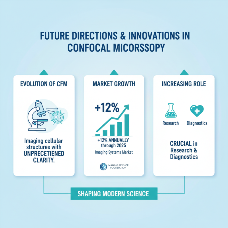
In the realm of contemporary scientific research, confocal fluorescence microscopy stands out as a pivotal tool. This technology enables researchers to visualize cellular structures with remarkable precision. Data from recent industry reports indicate that the global confocal microscopy market is projected to reach $3.29 billion by 2026. This growth underscores its significance across various fields, including biology, medicine, and material science.

Dr. Emily Tran, a leading expert in microscopy, aptly states, "Confocal fluorescence microscopy revolutionizes our understanding of complex biological processes." Her insight highlights the method's ability to provide high-resolution images, allowing scientists to explore cellular dynamics in real time. This level of detail was previously unattainable, fostering breakthroughs in drug development and disease research.

However, the reliance on confocal fluorescence microscopy is not without challenges. The technology requires extensive training and expertise. Additionally, issues related to phototoxicity and signal saturation can lead to distorted results. Researchers must continually refine their techniques and remain critical of their findings. The importance of confocal fluorescence microscopy in modern science is undeniable, yet it calls for an ongoing dialogue about its limitations and potential improvements.

Why Is Confocal Fluorescence Microscopy Important in Modern Science?

The Basics of Confocal Fluorescence Microscopy

Why Is Confocal Fluorescence Microscopy Important in Modern Science?

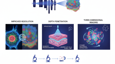
Confocal fluorescence microscopy is a pivotal tool in modern scientific research. This technique allows for high-resolution imaging of specimens at various depths. By using a laser to illuminate a sample, it captures detailed images while minimizing background noise. It achieves this through pinhole aperture technology, which enhances image contrast and clarity. According to a report by MarketsandMarkets, the global market for fluorescence microscopy is projected to reach $4.4 billion by 2025, indicating its growing significance.

This method has transformed fields like biology and materials science. In cellular biology, for instance, researchers can visualize live cells in real time. Enhanced imaging leads to improved understanding of cellular processes, which can impact medical research greatly. However, mastering this technology proficiently can take time. Users may face challenges with calibration and sample preparation.

**Tip:** Always optimize the pinhole size and settings for the best results.

The technology isn't without its limitations. Some samples might still produce background fluorescence, leading to potential misinterpretations. Researchers must remain vigilant in validating their findings. Rigorous controls are essential for accurate results.

**Tip:** Regularly check your microscope's calibration to ensure reliability.

Adjusting to the nuances of confocal systems can help in obtaining clearer images. As this field evolves, continuing education and practice remain paramount for researchers.

Historical Development and Evolution of the Technique

Confocal fluorescence microscopy has undergone significant advancements since its inception in the 20th century. Initially proposed by Marvin Minsky in 1955, the technique was a novel approach aimed at improving optical resolution. Early iterations faced challenges such as limited sensitivity and cumbersome design. Over the years, researchers refined the equipment, enhancing the ability to visualize biological specimens with greater clarity and detail.

The evolution of laser technology in the 1980s played a pivotal role. Lasers provided coherent light, allowing for deeper tissue penetration. This advancement revolutionized imaging techniques, extending the applicability of confocal microscopy in various fields. Yet, the increasing complexity of modern systems can pose challenges for new users. Mastery of this technology requires practice and experimentation, which can lead to initial frustration.

Despite its advancements, confocal fluorescence microscopy is not without limitations. The requirement for fluorescent labeling can introduce artifacts. Additionally, some samples may not emit fluorescence efficiently. Researchers must address these issues carefully to ensure reliable results. Continuous development is crucial as scientists push the boundaries of this powerful imaging tool.

Why Is Confocal Fluorescence Microscopy Important in Modern Science? - Historical Development and Evolution of the Technique

Year Milestone Significance Impact on Research
1955 First Confocal Setup Introduction of point-scanning technique Foundation for high-resolution imaging
1980 Commercialization First commercial confocal microscope Widespread use in biological research
1990s Injection of Laser Technology Enhanced imaging depth and speed Advanced cellular imaging techniques
2000s Multi-Photon Microscopy Reduced photodamage and deeper tissue imaging Expanded applications in vivo
Present Integration with Artificial Intelligence Automated image analysis and processing Enhanced research capabilities and efficiency

Key Applications in Biological and Medical Research

Confocal fluorescence microscopy plays a crucial role in biological and medical research. This advanced imaging technique allows scientists to visualize structures within cells with outstanding clarity. By using laser light, it enhances the contrast of fluorescent samples. Researchers can study complex cellular environments in real time. This is particularly valuable for understanding diseases at the cellular level.

One key application is in cancer research. Researchers can track the behavior of cancer cells as they invade surrounding tissues. This helps in understanding metastasis, a major challenge in cancer treatment.

Another notable application is in neuroscience. Scientists use confocal microscopy to visualize neural connections. This provides insights into brain function and disorders. However, the technique does have limits. It requires fluorescent labeling, which may alter cell behavior. Researchers must carefully consider these factors in their studies.

Advantages Over Traditional Microscopy Methods

Confocal fluorescence microscopy has revolutionized scientific imaging. It offers significant advantages over traditional microscopy methods. For instance, it provides high-resolution images. This technology uses a focused laser beam to illuminate samples. The result is the ability to capture fine details within cellular structures that other methods may miss.

Another important benefit is improved depth penetration. Confocal microscopy allows scientists to examine thicker specimens. They can visualize multiple layers of tissue in a single image. This is crucial for developmental biology and medical research. Yet, this method requires technical expertise. Not all laboratories have access to such advanced equipment.

Moreover, confocal microscopy minimizes background noise. This results in clearer images, enhancing accuracy in analyses. However, learning to interpret these images can be challenging. Researchers must often invest time in training. Despite these hurdles, the impact of this technique in modern science is undeniable. It opens up new avenues for exploration and discovery, pushing boundaries in various fields.

Advantages of Confocal Fluorescence Microscopy Over Traditional Microscopy

Confocal fluorescence microscopy offers significant advantages over traditional microscopy methods, including higher resolution, improved imaging depth, reduced background noise, the ability to create 3D images, and faster imaging speeds. These benefits make it a vital tool in modern scientific research.

Future Directions and Innovations in Confocal Microscopy

Why Is Confocal Fluorescence Microscopy Important in Modern Science?

Confocal fluorescence microscopy (CFM) continues to evolve, shaping the landscape of modern science. The ongoing innovations in this field bring new opportunities for imaging cellular structures with unprecedented clarity. As reported by the Imaging Science Foundation, the market for imaging systems is projected to grow by approximately 12% annually through 2025. This rapid advancement highlights the increasing role of CFM in research and diagnostics.

Emerging techniques, like super-resolution microscopy, have gained traction. These technologies push the boundaries of spatial resolution, allowing scientists to observe molecular interactions in real time. A recent study in Nature Methods revealed that combining CFM with advanced computational algorithms can enhance contrast and resolution, enabling deeper insights into cellular dynamics.

Tip: To achieve optimal results with confocal microscopy, ensure proper sample preparation. Poorly prepared samples can lead to artifacts that obscure true biological signals. Regular calibration of equipment also boosts reliability and reduces variability in imaging outcomes.

Despite the promise of CFM, challenges persist. Interpreting complex images can overwhelm researchers. Additionally, integrating CFM data with other imaging modalities requires careful planning and expertise.

Tip: Collaborate with specialists in data analysis. They can help navigate the intricacies of interpreting confocal images, ensuring that the insights drawn are both valid and actionable. The path forward for confocal fluorescence microscopy will rely on addressing these hurdles while embracing newfound innovations.

Search engine powered by ElasticSuite