Top 10 Applications of Confocal Imaging in Scientific Research?

Confocal imaging has revolutionized scientific research by providing detailed insights into cellular structures. Dr. Maria Chen, a leading expert in the field, once remarked, “Confocal imaging allows us to see what we couldn’t visualize before.” This technology enables researchers to obtain high-resolution images of samples, enhancing our understanding of intricate biological systems.

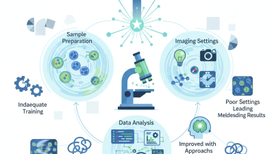
One of the remarkable aspects of confocal imaging is its versatility. It finds applications in various fields, such as biology, neuroscience, and materials science. In fluorescence microscopy, it sharpens the focus on specific targets within complex specimens. However, despite its advantages, there are challenges, including high costs and technical complexities.

Through the lens of confocal imaging, researchers can explore cellular dynamics and tissue architecture with unprecedented clarity. This imaging technique encourages us to reflect on our limitations. Achieving excellence in this field requires continual adaptation and learning. The future of confocal imaging looks promising, yet it also demands thoughtful consideration of its evolving role in scientific discoveries.

Top 10 Applications of Confocal Imaging in Scientific Research?

Applications of Confocal Imaging in Cell Biology and Tissue Analysis

Top 10 Applications of Confocal Imaging in Scientific Research?

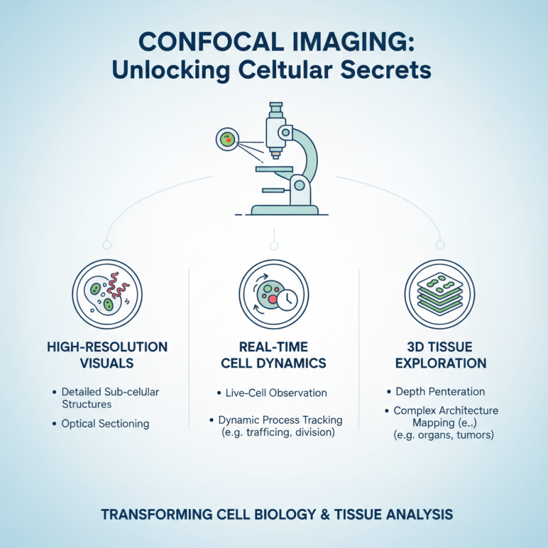
Confocal imaging has transformed our understanding of cell biology and tissue analysis. This technique provides high-resolution images. Scientists can visualize cells in real-time. It allows for the study of cellular processes at an unprecedented level. The depth of image capture enables researchers to explore complex tissues in 3D.

In cell biology, confocal imaging helps identify specific proteins within cells. Researchers mark these proteins with fluorescent tags. These tags light up under the confocal microscope. This method reveals detailed locations and interactions of proteins. However, challenges remain. Fluorescent tags can sometimes produce background noise. This noise may obscure vital information from the images.

For tissue analysis, confocal imaging allows examination of thin tissue sections. It captures detailed structures and functions. This technique is crucial in studying diseases like cancer. It helps in understanding how tumors develop. Yet, quantifying results can be problematic. Not all images are as clear as expected. Scientists must be diligent in interpreting their findings.

Enhancing Drug Discovery: Confocal Imaging in Pharmacology Research

Confocal imaging has become a vital tool in pharmacology research. This technology allows scientists to visualize and study cells and tissues at a microscopic level. By using lasers, researchers achieve high-resolution images. These detailed images reveal how potential drugs interact with biological components. This insight accelerates the drug discovery process.

However, confocal imaging is not without challenges. Image interpretation can be complex. Artifacts or noise may obscure the real data. These issues can mislead researchers about drug efficacy. Continuous refinement of imaging techniques is essential. Training researchers to analyze images with precision is also important.

Moreover, confocal imaging can be resource-intensive. The equipment requires significant investment. Access to this technology may not be available in all labs. This creates disparities in research opportunities. Effective collaboration and resource sharing could help bridge the gap. Ultimately, confocal imaging holds immense promise for advancing pharmacology research and drug development.

Applications of Confocal Imaging in Neuroscience: Revealing Brain Structures

Confocal imaging has transformed the field of neuroscience. Researchers can visualize brain structures with exceptional clarity. This technique uses laser light to scan samples, allowing scientists to capture detailed images of neurons. The precision enhances the understanding of brain connectivity and function.

By examining brain tissue slices, confocal imaging reveals intricate neuron networks. It helps identify the locations of synapses and the distribution of neurotransmitters. Such detailed insights can illuminate how brain circuits operate. However, this method does have limitations. For instance, thick samples might lead to light scattering, affecting image quality.

Moreover, confocal imaging requires meticulous sample preparation. It can be time-consuming and may introduce artifacts into results. Researchers must balance detail with efficiency. Despite these challenges, confocal imaging represents a powerful tool in neuroscience. It continues to unveil the complexities of the brain, fostering advancements in research and therapeutic strategies.

Utilizing Confocal Imaging for Multicolor Fluorescence in Molecular Biology

Confocal imaging has transformed molecular biology, particularly in multicolor fluorescence applications. This technique allows scientists to visualize multiple fluorescent markers in a single sample. By selectively illuminating thin sections of specimens, confocal imaging reduces background noise and enhances image clarity.

In studies of cellular interactions, researchers can employ various fluorescent dyes to tag different proteins. Each dye responds to specific wavelengths, making it possible to observe multiple processes simultaneously. For example, tracking the movement of two proteins within a living cell provides critical insights. However, challenges exist. Overlap in emission wavelengths can blur distinct signals, complicating interpretation.

Experimental design is crucial for success. Optimizing dye concentrations is essential; too much dye can lead to saturation. Moreover, careful planning around imaging settings will yield better results. Some findings may still surprise us. Images may not always match our expectations. Reflecting on these moments can inspire new approaches and questions. Clarity often arises from the imperfections we encounter in the lab.

Top 10 Applications of Confocal Imaging in Scientific Research

Advancements in Confocal Microscopy for Developmental Biology Studies

Confocal microscopy has transformed developmental biology studies. This technique allows researchers to capture images at various depths in a sample. High-resolution imaging reveals intricate cellular structures in embryos and tissues. By focusing on specific planes, scientists avoid the blurriness of traditional microscopy. This clarity uncovers vital processes like cell division and differentiation.

Tips for effective confocal imaging: Always calibrate your equipment before starting. Even slight misalignments can lead to poor image quality. Additionally, consider the sample's thickness. Sometimes, thicker samples may scatter light, complicating your results.

Another exciting advancement is live-cell imaging. This allows monitoring of dynamic processes in real-time. Scientists can watch how cells interact, move, and change. It opens doors to understanding complex biological systems. However, challenges still exist. Maintaining sample viability during imaging can be difficult. Researchers must find the right balance between observation and the health of the sample stored in culture.

Top 10 Applications of Confocal Imaging in Scientific Research

Application Field Description Year Introduced
Cell Structure Analysis Developmental Biology Detailed imaging of cellular components in living organisms. 1996
Neuron Visualization Neuroscience Studying the morphology of neurons and synaptic connections. 2001
Live Cell Imaging Cell Biology Monitoring dynamic processes in live cells over time. 2005
Fluorescence Localization Biochemistry Precise location of fluorescent tags on proteins. 2002
Tissue Imaging Histology Three-dimensional organization of tissue architecture. 2007
Drug Delivery Studies Pharmacology Analyzing the effectiveness of drug delivery systems. 2010
Gene Expression Analysis Genetics Visualizing localization and levels of gene expression. 2003
Pathogen Studies Microbiology Understanding the interactions between pathogens and host cells. 2015
Materials Science Materials Science Investigating the microstructural properties of materials. 2018
Environmental Studies Ecology Assessing biological responses to environmental changes. 2020
Search engine powered by ElasticSuite