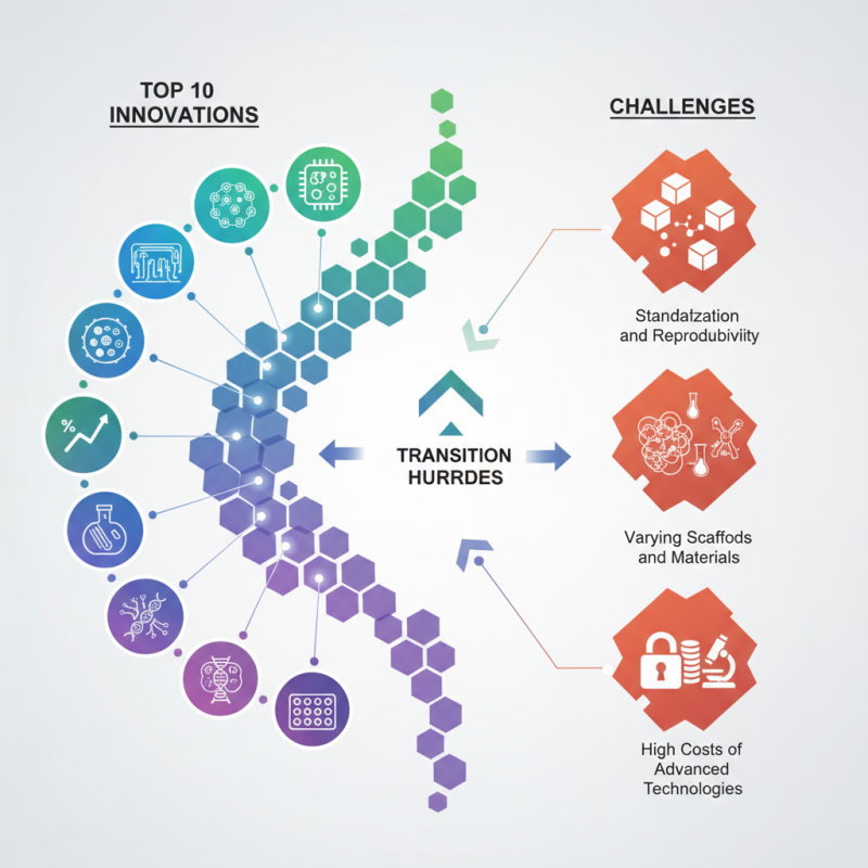
In recent years, the field of 3D cell culture has seen remarkable innovations, transforming the landscape of biomedical research. Data from the Global 3D Cell Culture Market report indicates a growth rate of 15% annually, highlighting its increasing significance. This technique enhances the physiological relevance of cellular behavior compared to traditional 2D cultures. Dr. Emily Carter, a leading expert in cellular biology, emphasizes, "3D cell culture models are essential for understanding complex tissue interactions."
However, the transition to 3D cell culture is not without challenges. Researchers often face difficulties in standardization and reproducibility. The varying scaffolds and materials can influence results significantly. Furthermore, the costs associated with advanced technologies can be a barrier for many laboratories. As the industry evolves, addressing these imperfections will be crucial for full integration into mainstream research practices. Exploring both the advancements and hurdles in 3D cell culture will unveil a deeper understanding of its role in innovative therapies.
In recent years, 3D cell culture technologies have transformed biomedical research. These advancements allow scientists to create more realistic models of human tissues. Traditional 2D culture methods often fail to replicate the complexities of real tissues. Now, researchers can study cell interactions in a more relevant environment. This is vital for drug testing and disease modeling.
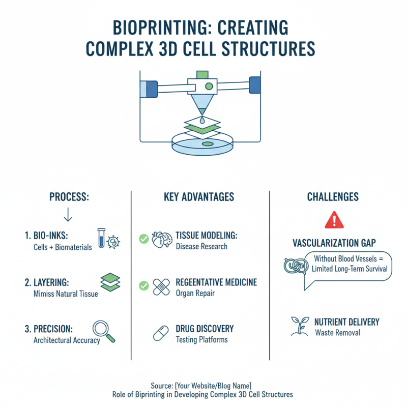
One exciting innovation is using bioprinting techniques to produce complex tissue structures. These structures mimic the architecture of human organs. Another remarkable advancement involves using hydrogels. Hydrogels provide a support system for cells, enhancing their growth and function. This improvement allows for better modeling of disease. Scientists can observe how cells react to treatments in a more accurate way.
Tip: Consider the scalability of different methods in your research. Smaller models can be easier to manage, yet larger ones might provide greater insights. Remember, not every method fits every research question. It's crucial to choose wisely based on your specific goals. Transparency in reporting your findings will also benefit the scientific community. Be sure to note any limitations in your model systems. This reflection helps in understanding their relevance.
This chart displays the impact scores of the top 10 innovations in 3D cell culture technologies. Each innovation is scored based on its significance and contribution to advancements in the field, with higher scores indicating greater impact.
The advancement of 3D cell culture systems relies heavily on the materials used. These materials provide a scaffold that mimics the natural environment of cells. Hydrogel-based materials are a popular choice. They can absorb water and have properties similar to natural tissue. This makes them ideal for supporting cell growth.
Another promising approach involves the use of natural polymers. They can be derived from sources like collagen or gelatin. These materials often enhance cell adhesion and proliferation. However, their variability can lead to inconsistent results. Researchers must carefully optimize conditions for each experiment.
Synthetic materials offer consistency. They can be designed for specific purposes and often have reproducible properties. Yet, they may lack the biological cues that promote natural cell behavior. Finding the right balance between natural and synthetic materials remains a challenge. This ongoing exploration pushes the boundaries of 3D cell culture innovation.
Bioprinting plays a crucial role in creating complex 3D cell structures. This technology allows scientists to layer cells, mimicking natural tissue architecture. By utilizing bio-inks made from cells and biomaterials, researchers can print living tissues with remarkable precision. However, challenges remain in achieving vascularization. Without blood vessels, tissues cannot thrive long-term.
Another fascinating aspect of bioprinting is the ability to design customized structures. Personalized tissues can be produced for drug testing or regenerative medicine. Yet, the development of functional tissues is still in its infancy. While we see promising results, issues like cell viability and integration need thorough exploration.
The potential for bioprinting is vast, but we must address these hurdles carefully. Each breakthrough brings hope, but often, experimentation leads to disappointment. Striking a balance between innovation and realistic expectations is essential for progress. Advancements will continue to shape how we approach regenerative medicine in the future.
In recent years, 3D cell culture has transformed our understanding of cellular behavior. Traditional 2D cultures often fail to mimic the complex environment of living tissues. According to a report by the National Institutes of Health, about 90% of drug candidates fail in clinical trials due to inadequate models. This highlights a significant need for improved methods in research.
Innovative techniques are emerging to monitor cell behavior in 3D settings. For example, live-cell imaging technologies allow researchers to observe real-time cellular interactions. A study showcased that 3D cultures could enhance cell signaling pathways, increasing the accuracy of drug responses. Yet, challenges remain. Many imaging methods struggle with resolution and depth, limiting their effectiveness in dense cultures.
Another notable advancement is the use of microfluidics to create more physiological conditions. This technique enables precise control over the microenvironment. Reports indicate that experiments using microfluidic devices yield a 40% higher data reliability in studying tissue responses. However, difficulties in scaling these technologies for larger studies persist. Researchers must address these hurdles to fully realize the potential of 3D cell culture. Exploring these innovations could reshape therapeutic development in significant ways.
| Innovation | Description | Applications | Advantages |
|---|---|---|---|
| Bioprinting | A method that uses 3D printing technology to create living tissues. | Tissue engineering, regenerative medicine. | Precision, scalability, and customization of tissue structures. |
| Microfluidics | Manipulation of fluids at the microscale for creating 3D microenvironments. | Drug testing, disease modeling. | Reduced sample size, real-time monitoring. |
| Hydrogel scaffolds | Gelatinous materials that provide a supportive environment for cell growth. | Cell culture, regenerative therapies. | Biocompatibility, tunable properties. |
| Organoids | 3D miniaturized organs derived from stem cells that mimic native organ structure. | Disease modeling, drug discovery. | In vivo-like behavior, complexity. |
| 3D Matrices | Structured environments that support cell adhesion, growth, and function. | Cancer research, personalized medicine. | Enhanced cell interaction, mimicking tissue architecture. |
| Live-cell Imaging | Techniques to visualize living cells in real time. | Cell behavior study, drug response observation. | Dynamic insights, temporal resolution. |
| Tissue-on-a-Chip | Microdevices that mimic the physiological responses of human organs. | Drug screening, toxicity testing. | High-throughput, reproducibility. |
| Automated Cell Culture | Use of robotics and automation to manage cell cultures. | Scale-up production, consistency in results. | Reduced human error, increased efficiency. |
| Bioreactors | Devices that provide a controlled environment for cell growth. | Tissue engineering, pharmaceuticals production. | Scalability, optimal growth conditions. |
| CRISPR for 3D Culture | Using gene editing technology to enhance 3D cell culture systems. | Genetic studies, disease modeling. | Precision modifications, tailored studies. |
3D cell culture is revolutionizing drug development and disease modeling. Unlike traditional 2D cultures, 3D systems mimic the natural environment of human tissues. This allows researchers to study complex cellular interactions in a more realistic manner. In drug development, 3D cultures can lead to more accurate predictions of drug efficacy and safety.
One notable application is in cancer research. Researchers can use 3D cell cultures to observe tumor growth and behavior. By doing so, they can identify effective therapies. This approach enhances the understanding of drug resistance mechanisms. The results are more relevant than those from flat cultures.
Tip: When designing a 3D culture experiment, focus on the specific properties of the cells you are studying. Consider their growth patterns and interaction types.
Another area of focus is neurological disorders. 3D cultures can recreate brain-like structures. This helps in modeling conditions like Alzheimer’s disease. Understanding cellular pathways is crucial for developing targeted therapies. It’s important to refine the models for better accuracy.
Tip: Evaluate your model periodically. Does it truly represent the disease state? This reflection is key to advancing research.
In summary, 3D cell cultures show great promise. They can provide insights into drug responses and disease mechanisms. However, continuous improvement and evaluation of these systems are needed for optimal results.