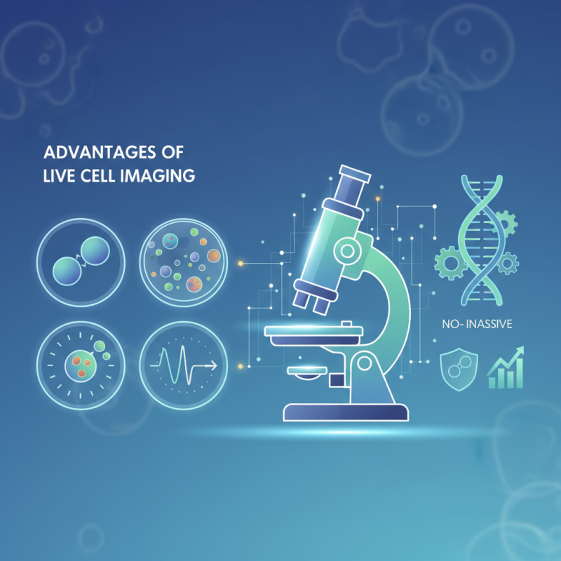
Live cell imaging microscopes have revolutionized biological research. They allow scientists to observe living cells in real-time. This technique provides insights into cellular processes that static imaging cannot reveal. Researchers can study cellular dynamics, including movement, division, and interactions.
One significant advantage is the ability to monitor cellular behavior without harming the cells. This non-invasive approach leads to more accurate data. Scientists can capture the effects of drugs over time, offering a clearer picture of cellular responses. Additionally, these microscopes support the analysis of complex biological systems.
Despite their benefits, challenges exist. Live cell imaging often requires specialized training and expertise. Moreover, equipment costs can be substantial. Researchers must consider these factors when integrating live cell imaging into their work. Nevertheless, the advantages are undeniable and can lead to breakthroughs in understanding life at the cellular level.
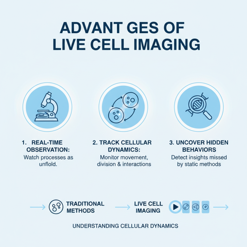
Live cell imaging has transformed our understanding of cellular dynamics. This technique allows researchers to observe living cells in real-time. By capturing cellular processes as they unfold, scientists can gain insights into complex behaviors and interactions. For instance, tracking cell division provides a direct view of mitotic phases. This real-time observation can reveal unexpected behaviors that traditional methods may miss.
The advantages extend beyond mere observation. Researchers can measure cellular responses to external stimuli. It allows for the visualization of drug effects on live cells. This knowledge can lead to better therapeutic approaches. However, the technology comes with challenges. Image clarity may vary due to cellular movement. Balancing resolution and speed remains a critical consideration. Over time, these limitations can push scientists to refine their techniques, fostering innovation. Exploring these dynamics reveals the intricate networks that govern life.
Enhanced temporal resolution in live cell imaging techniques plays a critical role in advancing biological research. The ability to capture dynamic cellular processes in real-time offers researchers unprecedented insights. According to a report from the National Institutes of Health, live cell imaging can achieve frame rates of up to 100 frames per second. This capability allows scientists to observe rapid cellular events, such as membrane trafficking and cellular signaling.
Despite these advancements, challenges remain. Achieving high temporal resolution sometimes compromises spatial resolution. This trade-off can lead to artifacts in the data. Researchers must carefully design their experiments to balance these parameters. Furthermore, capturing live cell images frequently requires intricate setups that can be costly and time-consuming.
Overcoming these obstacles is essential for maximizing the benefits of live cell imaging. Researchers are encouraged to engage in collaborative studies to share best practices. They can also utilize integrated approaches that combine multiple imaging techniques. This collaborative effort helps refine methodologies, leading to more reliable results in cellular biology. Ultimately, enhancing temporal resolution in live cell imaging techniques is a double-edged sword that requires ongoing reflection and adaptation in the research community.
Live cell imaging microscopy offers a transformative approach to understanding cellular processes in real-time. By allowing researchers to observe living cells without disrupting their natural environment, this technology provides insights that are unattainable with traditional microscopy. Researchers can monitor cellular behaviors, such as division, movement, and signaling, as they happen. This real-time observation is crucial for studying dynamic processes like cell differentiation and response to treatment.
The ability to visualize these processes can lead to breakthroughs in various fields, including cancer research and developmental biology. However, this technique is not without its challenges.
Maintaining cell health during prolonged imaging is often a concern. Environmental control, including temperature and CO2 levels, is essential but can be technically demanding. Moreover, data interpretation from live imaging requires expertise. The complexity of biological systems can lead to misinterpretation if care is not taken.
While live cell imaging opens new avenues for discovery, researchers must adapt to its intricacies. The balance between observation and maintaining cell integrity needs careful consideration. Innovations in imaging techniques continue to evolve, yet there is always room for improvement. Effective training and collaboration can enhance the reliability of findings from live cell imaging studies.
Live cell imaging is a powerful technique that has transformed biological research. However, one of the critical challenges it faces is phototoxicity, which can adversely affect cell viability. Phototoxicity occurs when fluorescent light used in microscopy causes damage to living cells. Research indicates that excessive light exposure can lead to up to a 70% reduction in cell viability. This emphasizes the importance of minimizing light exposure during experiments.
There are several methods to reduce phototoxicity in live cell imaging. First, using advanced light sources, like LED or lasers, can significantly lower the energy needed for imaging. Studies show that LEDs can reduce photobleaching by approximately 50% compared to traditional halogen lamps. Another strategy is to optimize imaging parameters, such as exposure time and intensity. Adjusting these factors can help maintain cell health without compromising the quality of the images captured.
Despite these strategies, researchers must remain cautious. Even with optimized conditions, phototoxic effects can still occur. Some cells are more sensitive than others, and factors like cell type and imaging duration play a crucial role. Understanding these nuances can help refine imaging protocols for better results. Continuous assessment and adaptation are necessary to ensure cell viability while obtaining high-resolution images.
This chart illustrates the top 10 advantages of live cell imaging microscopes for research, emphasizing how minimizing phototoxicity significantly enhances cell viability and overall research outcomes. Each advantage is rated based on its impact in live cell imaging applications.
Live cell imaging technology plays a critical role in drug discovery and development. It allows researchers to observe cellular processes in real-time. This methodology helps identify how cells respond to potential drugs. According to a report from the National Institutes of Health, nearly 70% of drug candidates fail in clinical trials due to poor efficacy or safety. Live cell imaging can improve success rates by providing insights into cellular reactions to therapies.
Specific applications in drug discovery include assessing drug efficacy. For instance, researchers can monitor changes in cell morphology and behavior. This continuous observation helps determine the optimal concentration of a drug. A study published in Nature Methods highlighted that real-time imaging can cut drug development time by 30%. This reduction correlates with savings of millions in research costs.
Despite its advantages, challenges exist. The imaging process can sometimes distort cellular activities. Artifacts may arise when cells are under observation for extended periods. Additionally, interpreting the data requires specialized skills. There is a learning curve to effectively analyze results. Researchers must balance the need for detail with the complexity of data collection. These reflections are essential for advancing drug discovery and enhancing the reliability of findings.
| Advantage | Description | Applications in Drug Discovery | Impact on Research |
|---|---|---|---|
| Real-Time Observation | Allows researchers to visualize biological processes as they occur. | Monitoring drug effects on living cells. | Enhances understanding of drug mechanisms. |
| Cell Viability Assessment | Evaluates the health of cells in real time. | Determining cytotoxic effects of compounds. | Improves safety profiles of drugs. |
| High-Throughput Screening | Facilitates analysis of multiple samples simultaneously. | Accelerating lead compound identification. | Increases efficiency in drug development. |
| Complex Interaction Studies | Enables detailed observation of cellular interactions. | Investigating tumor microenvironments. | Enhances comprehension of drug action in complex systems. |
| Longitudinal Studies | Allows observation over extended periods. | Tracking disease progression and therapy response. | Provides critical insights for treatment optimization. |
| Multicolor Imaging | Simultaneous visualization of multiple targets. | Examining signaling pathways | Enhances understanding of pharmacodynamics. |
| Quantitative Analysis | Facilitates precise measurements of cellular parameters. | Assessing drug efficiency quantitatively. | Supports robust data for regulatory submissions. |
| User-Friendly Interfaces | Modern software simplifies operation and data analysis. | Eases implementation in various laboratories. | Sets the stage for innovative researchers. |
| Therapeutic Target Validation | Validates the effectiveness of potential therapeutic targets. | Refining targets for drug development. | Increases success rates in clinical trials. |
| Customization for Specific Needs | Adaptable systems for varying research applications. | Tailoring imaging settings for unique experiments. | Maximizes productivity and relevance of findings. |