What Is the Best Confocal Imaging System to Buy in 2026?
Choosing the best confocal imaging system in 2026 begins with a less glamorous question: what must you see clearly? A live-cell lab may value gentle illumination and environmental control. A core facility may need fast scanning, flexible detectors, and reliable service. Thick tissue, fluorescent labels, and limited bench space each change the decision. There is no universal winner. That is inconvenient, but useful.
Confocal pioneer Marvin Minsky’s work points to a lasting principle: optical sectioning matters when out-of-focus light obscures the detail you need. Put plainly, buy for the specimen, not the feature list. Compare image quality on representative samples, not only polished vendor demonstrations. Check detector options, laser lines, objective compatibility, software workflow, training, and local support. Ask how the system handles photobleaching during long time-lapse sessions. Small details count. A crowded microscope room can make a large footprint a daily problem.
A careful shortlist should include the full cost of ownership, including maintenance, upgrades, and staff time. Request demonstrations with your own samples, then review signal-to-noise, acquisition speed, and repeatability. Also ask what the system cannot do well. Confocal imaging can produce crisp optical sections, but it does not remove every limit imposed by scattering, phototoxicity, or sample preparation. I would treat any single “best” recommendation with some caution. The best purchase is the one your team can use consistently, support locally, and justify with real data.
What Defines a Confocal Imaging System and How Does It Work?
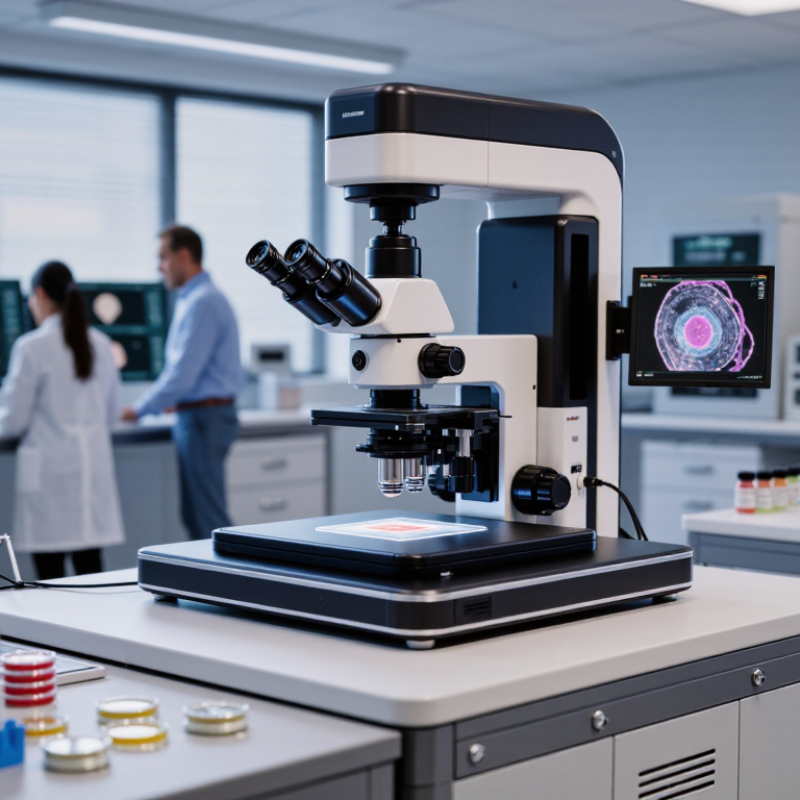
A confocal imaging system is a microscope that rejects much of the out-of-focus light. It uses focused illumination and a small aperture, called a pinhole, placed before the detector. Light from the focal plane passes through; blurred light from above and below is largely blocked. The result is an image with improved contrast, especially in thicker or layered samples. The pinhole is not magic. If it is set too small, the image can become dim, and longer exposure may increase bleaching.
In a typical setup, a laser scans the sample point by point, while detectors record emitted or reflected light. Software assembles those signals into an image, and changing the focal position can produce optical sections through a specimen. For example, a researcher can inspect fluorescent structures at several depths without physically slicing the sample. That detail matters. Resolution and image quality still depend on the objective, detector sensitivity, sample preparation, and careful alignment. In practical use, I would check for stable focus, manageable acquisition time, and clear controls for laser power and pinhole size. A confocal system can reveal useful detail, but it cannot restore information the sample never provided. Even experienced operators sometimes need to revisit settings when a bright surface hides dim features deeper inside.
What Is the Best Confocal Imaging System to Buy in 2026?
What Defines a Confocal Imaging System and How Does It Work?
Common excitation laser lines used in confocal microscopy are shown below. A confocal system scans focused laser light across a sample and uses a pinhole to reject out-of-focus light before detection, enabling optical sectioning. The best configuration depends on the fluorophores, sample, resolution, and imaging speed required.
Which Confocal Imaging Technologies Are Available in 2026?
In 2026, confocal imaging systems use several approaches to reject out-of-focus light. Point-scanning laser confocal microscopes illuminate one small spot at a time. They offer flexible optical sectioning and suit fixed samples or slower live-cell work. But scanning can be slow, and intense illumination may stress delicate specimens.
Small details matter.
Spinning-disk systems illuminate many points in parallel, making them useful for faster time-lapse imaging. They can reduce exposure per point, though thick or strongly scattering samples may produce less even results. Line-scanning confocal systems capture a line at once and can balance speed with optical sectioning for selected applications. Some platforms add spectral detectors, which separate overlapping fluorescent signals by their emission profiles. Detector sensitivity, pinhole settings, objective choice, and sample preparation all affect image quality. A system that looks impressive in a specification sheet may disappoint with your actual cells. That is worth checking.
For a purchase decision, compare image speed, usable depth, fluorescence channels, and maintenance needs using representative samples. Ask for raw image files, not just polished demonstrations. No single technology wins every experiment, and I would treat any simple ranking with caution.
How Do Research Needs Shape the Choice of Confocal System?
Research needs should drive the purchase, not a crowded feature list.
For thin, fixed cells, a standard point-scanning confocal may provide sufficient contrast and optical sectioning. Thick organoids or cleared tissue demand stronger signal collection and careful attention to imaging depth. Live-cell experiments need fast acquisition, stable temperature and gas control, and low light exposure. Otherwise, the cells may change while the system captures them.
Start with the samples.
How many colors must be separated? How quickly do they move? What is the largest field you need to capture? A spectral detector can help distinguish overlapping emission, while a resonant scanner can reduce capture time. Neither solves every problem. More laser power may improve signal, but it can also increase photobleaching and phototoxicity. Small details matter.
Budget planning should include objectives, detectors, service, and training—not just the microscope body. Grand View Research’s 2024 Confocal Microscope Market report estimated the global market at USD 1.1 billion in 2023 and projected 5.6% annual growth through 2030. That signals expanding adoption, not a universal best choice. Before buying, test representative samples and compare usable image quality, acquisition time, and operating costs. A polished demo can still miss your real workflow.
Which Performance, Compatibility, and Cost Factors Should Buyers Compare?
A confocal system should match the samples you actually image, not just the most impressive specification sheet. Compare lateral and axial resolution, detector sensitivity, and scanning speed using representative samples. A dim fluorescent signal may need sensitive detection more than rapid scanning. Bright, moving specimens may need the reverse. Ask for a demonstration with your own slides, including a thick sample if that is routine.
Check compatibility before discussing price. Confirm that the system supports your fluorophores, excitation wavelengths, emission channels, objectives, and existing microscope components. Verify that image files can be exported in formats your analysis software can read. Small details matter: a filter set that misses an emission peak can weaken useful signal. So can software that makes routine measurements awkward. Compatibility lists can be incomplete, so get key requirements in writing.
Compare the full cost of ownership, not only the purchase quote. Include installation, service coverage, staff training, computer upgrades, and any needed objectives or environmental controls. Request expected maintenance costs and typical repair timelines. Downtime has a cost too. A lower-priced system may be sensible for occasional imaging, while a heavily used facility may value local service and easier training. Still, estimates rarely capture every workflow snag; leave room in the budget for an imperfect first year.
How Can Buyers Evaluate Confocal Imaging Systems for Different Applications?
Evaluate a confocal imaging system against the samples you actually study, not only a polished demonstration slide. For thin, fixed cells, resolution and reliable fluorescence separation may matter most. For living cells, check whether the instrument can capture useful images with low light exposure. A bright image is not always a healthy sample.
For thick tissue, ask how well the system rejects out-of-focus light and whether its working distance suits your objectives. Look closely at detector sensitivity, available excitation wavelengths, and the practical range of optical sectioning. If you image moving samples, test acquisition speed with your real settings. A fast scan that produces noisy data may not help.
Request a hands-on trial using representative samples. Note the time needed to set up an experiment, align the system, and export usable files. Ask about training, service access, software updates, and compatibility with your analysis workflow. Small details matter. A menu that feels confusing during a short demo may slow daily work. Compare total operating costs, including maintenance and consumables, rather than purchase price alone. I would not treat any single resolution figure as decisive; sample preparation and operator skill can change results substantially. Some compromises remain. Decide which limitations your lab can live with.