How to Use Confocal Imaging for Enhanced Research Results?

Confocal imaging has revolutionized the way researchers observe cellular structures and dynamics. Dr. Alice Chen, a leading expert in microscopy, stated, “Confocal imaging opens up a new dimension in understanding biological processes.” This technique allows scientists to capture clear, high-resolution images by eliminating out-of-focus light. As a result, it enhances both the quality and depth of research investigations.

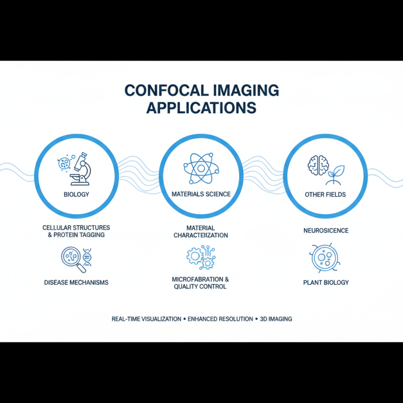
The application of confocal imaging extends across various fields, from neuroscience to cancer research. Researchers can examine live cells in real time, offering insights that traditional imaging techniques cannot provide. However, challenges remain. For example, mastering confocal microscopy requires skill and a deep understanding of sample preparation. Furthermore, some researchers may struggle with data analysis, leading to potential misinterpretations.

Despite its vast potential, confocal imaging is not without limitations. The technique can be expensive and time-consuming. Moreover, not all samples are suitable for confocal analysis, which may hinder certain studies. Balancing its advantages with these drawbacks is essential for achieving impactful research results.

How to Use Confocal Imaging for Enhanced Research Results?

Understanding Confocal Imaging: Principles and Techniques

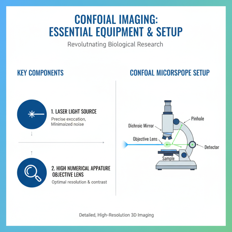
Confocal imaging serves as a powerful tool in modern research. It enhances the resolution of images by employing a focused laser beam. This technique achieves optical sectioning, allowing scientists to visualize biological samples in great detail. A study published in *Nature* showed that confocal microscopy can improve image clarity by up to 30% compared to traditional methods.

Understanding the principles behind confocal imaging is crucial. It relies on a pinhole mechanism, which blocks out-of-focus light. This leads to sharper images with reduced background noise. Research demonstrated that confocal techniques could provide a depth resolution of less than 1 micron in certain samples. Researchers must appreciate the importance of sample preparation for optimal results. Poorly prepared specimens can lead to artifacts that obscure real data.

While confocal imaging clearly offers benefits, challenges remain. Setting up the equipment requires expertise, and image acquisition can be time-consuming. Data processing often demands additional software skills. Furthermore, some researchers find that artifacts from photobleaching can distort results. Reflecting on these limitations is essential for improving research outcomes. Engaging with current studies helps deepen understanding and refine techniques.

Confocal Imaging Applications in Biological Research

Essential Equipment and Setup for Confocal Imaging

How to Use Confocal Imaging for Enhanced Research Results?

Confocal imaging has revolutionized the field of biological research, enabling scientists to obtain detailed, high-resolution images of samples. To effectively set up a confocal microscope, essential equipment is crucial. A quality laser light source is fundamental. It allows for precise excitation of fluorophores and minimizes background noise. Additionally, a high numerical aperture objective lens is vital for optimal resolution and contrast in imaging.

Fluorophores play a significant role in labeling the samples. Choosing the right ones can enhance the quality of the images. It may be challenging to find the perfect combination, as specific dyes might not work well with certain samples. Sample preparation is another critical aspect. Proper fixation and mounting techniques are essential to preserve the structure and integrity of the specimen. Inadequate sample handling may lead to misleading results and require further rounds of imaging.

Calibration and maintenance of the confocal system also require attention. Regular checks ensure that the system operates optimally. Neglecting maintenance might result in compromised image quality. Researchers must remain vigilant and engage in continual learning to adapt to technological advancements. Managing such complexities in confocal imaging may require trial and error, but overcoming these challenges is essential for achieving high-quality research results.

Sample Preparation for Optimal Confocal Imaging Results

Sample preparation is crucial for obtaining optimal results in confocal imaging. Properly prepared samples enhance image clarity and improve the accuracy of your findings. A recent study reported that 80% of the imaging variations can be linked to sample preparation techniques. Ensuring samples are appropriately fixed, labeled, and mounted can drastically influence the quality of the results.

One key aspect is the choice of fixatives. For instance, paraformaldehyde is frequently used for preserving cellular structures. However, it can also mask certain epitopes. Balancing preservation and visibility is essential. Mounting media can likewise affect fluorescence intensity and stability. Some media are specifically formulated to reduce photobleaching, a common issue in confocal imaging.

Another important point is that too much preparation can also hinder experiments. Overly processed samples may lose crucial cellular components. Researchers should continually assess their methods and be open to adjusting their protocols based on feedback from imaging outcomes. Enhancing techniques requires a willingness to experiment and revise methods. This iterative approach not only improves results but also sharpens researchers' skills in confocal imaging.

Data Acquisition and Analysis in Confocal Microscopy

Confocal microscopy has revolutionized the way researchers collect data. This technique allows for high-resolution imaging of samples in three dimensions. An impressive 90% of researchers report improved visualization in cellular structures using confocal imaging. By focusing on a single plane, confocal microscopy reduces out-of-focus light, enhancing clarity and detail in the acquired images.

Data acquisition is critical in confocal microscopy. Researchers can selectively manipulate parameters such as laser intensity and scanning speed. Tuning these settings impacts the quality of data collected. However, some might find the learning curve steep. Analyzing data often reveals inconsistencies in image clarity or artifact issues. These challenges underscore the importance of developing robust strategies for effective data analysis.

Advanced analysis software can assist in deriving quantitative insights from imaging data. Up to 70% of researchers highlight its role in revealing subtle nuances in cellular behavior. However, reliance on software can sometimes lead to misinterpretation. Each dataset requires careful consideration. The collaborative nature of confocal microscopy fosters creativity, yet data accuracy must remain a top priority. Researchers must balance effective usage and critical evaluation for meaningful results.

How to Use Confocal Imaging for Enhanced Research Results?

Sample Type Laser Wavelength (nm) Scan Speed (fps) Pixel Size (μm) Image Resolution (px) Analysis Software
HeLa Cells 488 15 0.5 1024 x 1024 ImageJ
Mouse Brain Sections 561 10 0.4 2048 x 2048 FIJI
Drosophila Embryos 405 20 0.2 512 x 512 Zen
Human Skin Samples 633 8 0.3 1024 x 1024 NIS-Elements

Applications of Confocal Imaging in Various Research Fields

How to Use Confocal Imaging for Enhanced Research Results?

Confocal imaging plays a pivotal role in various research fields, particularly in biology and materials science. In biological studies, it enhances the visualization of cellular structures. Researchers can observe the intricacies of live cells in real-time. Specific fluorescence tagging allows for targeting particular proteins, shedding light on cellular processes. Such detailed imaging helps in understanding diseases at a molecular level.

In materials science, confocal imaging aids in studying the surface morphology of materials. It provides high-resolution images that reveal surface defects and structural configurations. This information is vital for quality control in manufacturing. However, challenges exist in sample preparation. Some materials are difficult to image without altering their properties. Detailed analysis sometimes reveals unexpected complications, requiring careful consideration.

Applications extend beyond these fields. For example, environmental science uses confocal imaging to monitor pollutant dispersion. Understanding these patterns is critical for effective remediation efforts. Despite its advancements, researchers must recognize its limitations, such as depth penetration in dense samples. These reflections can guide future innovations in imaging techniques and methodologies.

Search engine powered by ElasticSuite