
How to Choose Trypsin EDTA for Cell Culture in 2026?
Choosing trypsin EDTA in 2026 requires more than comparing concentration, package size, or price. Cell lines respond differently to detachment stress. Primary cells may round quickly, while robust immortalized cells can tolerate longer exposure. The wrong formulation can reduce viability, alter morphology, and compromise downstream assays. Small details matter. A warm bottle, repeated pipetting, or delayed neutralization may change the result.
Industry reports show continued growth in cell culture, biopharmaceutical manufacturing, and regenerative medicine. Grand View Research highlights expanding demand for reliable cell-based workflows. MarketsandMarkets also identifies cell culture media and related reagents as essential segments in bioprocessing. These reports support a practical point: trypsin EDTA is a routine reagent, but its performance affects valuable experiments. Product selection should consider EDTA strength, trypsin activity, animal-origin status, sterility, traceability, and compatibility with the target cells.
R. Ian Freshney, author of Culture of Animal Cells, is often quoted saying, “Cell culture is an art as well as a science.” That warning remains relevant. A certificate of analysis cannot replace controlled handling and documented recovery rates. Compare products under your own laboratory conditions. Measure detachment time, post-passage viability, attachment quality, and cell morphology. Do not rely on marketing language alone. A cleaner label is not automatically a better choice. Some recommendations will still need revision after real-world testing. That is the uncomfortable part. The best trypsin EDTA is not universally superior; it is the formulation that consistently protects your cells and supports your specific workflow.
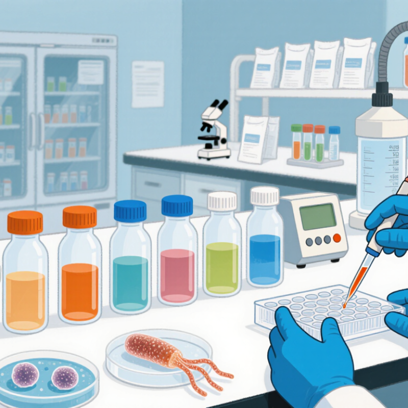
Understanding Trypsin-EDTA Composition and Cell Detachment Principles
How to Choose Trypsin EDTA for Cell Culture in 2026?
Trypsin-EDTA is not a single chemical. It combines trypsin, a serine protease, with EDTA, a calcium and magnesium chelator. A common working formula contains 0.05% trypsin and 0.02% EDTA. Exact concentrations vary by cell type and supplier formulation. The 2024 Global Cell Culture Market Report identified consumables as a major market segment, with double-digit growth projected through 2030. That growth reflects wider use, not automatic suitability.
Trypsin cuts adhesion proteins near the cell surface. EDTA removes divalent ions that support cell-cell and cell-surface attachment. Together, they loosen adherent cells for collection. Under a microscope, suitable detachment often appears as rounded cells with bright edges. Most cells release within two to five minutes at room temperature. Timing matters. Overexposure can reduce membrane integrity and recovery. Gentle tapping may help, but forceful pipetting can create visible debris.
In practice, I match the formulation to the cell line, passage number, and culture surface. Primary cells usually need milder exposure than robust immortalized lines. I also check pH, sterility, storage temperature, and documented activity. A 2023 laboratory workflow survey reported that inconsistent detachment timing remained a frequent source of cell-count variation. My own judgment can still fail here. A familiar protocol may not suit a new matrix. Test a small culture first, record the exposure time, and compare viability after reseeding.
How to Choose Trypsin EDTA for Cell Culture in 2026? - Understanding Trypsin-EDTA Composition and Cell Detachment Principles
| Selection Dimension | Typical Value or Option | Cell Detachment Principle | Practical Selection Guidance |
|---|---|---|---|
| Trypsin concentration | 0.05% or 0.25% w/v are common working formulations | Trypsin is a serine protease that cleaves proteins involved in cell–cell and cell–substrate adhesion. | Use a lower concentration for sensitive or easily detached cells; a higher concentration may be useful for strongly adherent cultures, provided exposure is controlled. |
| EDTA concentration | Approximately 0.02% w/v, equivalent to about 0.5 mM when disodium EDTA dihydrate is used | EDTA chelates calcium and magnesium ions that support integrin-mediated adhesion and cell–cell contacts. | Choose EDTA-containing formulations when divalent-cation-dependent adhesion needs to be weakened before proteolysis. |
| Trypsin-to-EDTA balance | Low trypsin/EDTA: gentler action; higher trypsin/EDTA: faster and stronger detachment | EDTA first reduces ion-dependent adhesion, allowing trypsin to access adhesion-associated proteins more efficiently. | Optimize the combination rather than increasing trypsin alone when cell-surface preservation is important. |
| Calcium and magnesium content | Prefer Ca2+/Mg2+-free diluent for standard detachment workflows | Removing divalent cations reduces adhesion and prevents them from counteracting EDTA chelation. | Check the formulation and rinse solution carefully; residual calcium or magnesium can reduce detachment efficiency. |
| pH range | Usually near physiological pH, approximately 7.2–7.8 | Trypsin activity and cell tolerance are both influenced by pH. | Use a formulation validated for mammalian cell culture and avoid prolonged exposure outside the recommended pH range. |
| Exposure temperature | Room temperature or approximately 37°C, depending on the cell line and protocol | Higher temperature generally accelerates enzymatic activity and detachment. | Use room temperature for slower, more controllable action; use 37°C only when validated for the specific culture. |
| Expected exposure time | Often about 2–10 minutes, but cell-line dependent | Detachment time depends on adhesion strength, confluence, matrix coating, temperature, and reagent activity. | Monitor the culture microscopically and neutralize or remove the reagent as soon as cells detach. |
| Serum compatibility | Serum-containing medium can inhibit trypsin through protease inhibitors | Serum proteins reduce residual protease activity after cell release. | Use complete growth medium or another validated inhibitor solution promptly after detachment. |
| Cell-surface sensitivity | Sensitive cells require reduced protease exposure; robust adherent cells may tolerate stronger treatment | Excessive proteolysis can remove or alter surface proteins and may reduce viability or recovery. | Start with the mildest formulation that achieves consistent detachment and minimize incubation time. |
| Culture format | Standard tissue-culture plastic, coated plates, multilayer vessels, or microcarriers | Surface chemistry and extracellular-matrix coatings change adhesion strength and reagent access. | Expect longer or stronger treatment on coated and three-dimensional systems; validate each format separately. |
| Cell density and confluence | Common subculture range: approximately 70–90% confluence, depending on the cell type | Overconfluent cultures may develop stronger cell–cell contacts and altered morphology. | Subculture within the cell line’s validated confluence window for more reproducible detachment. |
| Sterility and storage | Use sterile, cell-culture-grade material; follow the supplier’s temperature and freeze–thaw limits | Repeated warming and freeze–thaw cycles can reduce enzyme performance and increase variability. | Use aliquots when appropriate, avoid unnecessary temperature cycling, and discard solutions showing contamination or unexplained performance changes. |
Related Posts
-
What Is the Best Trypsin EDTA Supplier in China?
-

2026 Top Uses of Paxgene Blood RNA Tubes in Medical Research?
-
2026 How to Choose the Best Polyclonal Antibody for Research?
-

What Is a Polyclonal Antibody and What Types Are Available?
-

2026 How to Use a Live Cell Imaging Microscope Effectively?
-
What is a cas9 antibody and how is it used?