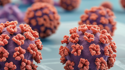
A mycoplasma detection kit helps laboratories identify tiny bacteria that can contaminate cell cultures, bioprocessing materials, and biological products. Mycoplasmas lack traditional cell walls, making them difficult to observe with routine staining. They may also grow slowly, while quietly changing cell growth, metabolism, and experimental results.
Most kits use molecular detection, especially polymerase chain reaction (PCR) or quantitative PCR. A technician collects a sample, releases its genetic material, and adds prepared reagents. If mycoplasma DNA is present, selected genetic regions are amplified. The instrument then records fluorescence or another measurable signal. Stronger signals usually suggest more target DNA, although interpretation depends on the kit’s design and controls.
Controls matter.
A reliable workflow includes positive, negative, and extraction controls. These checks help reveal reagent failure, contamination, or sample inhibition. From practical laboratory experience, careful pipetting and separated work areas often matter as much as the instrument. A clean-looking result is not automatically a trustworthy result. No kit is perfect. Some organisms may escape detection if the target sequence does not match, the sample contains inhibitors, or the contamination level is very low. For that reason, trained personnel should follow validated instructions, document sample handling, and confirm unexpected findings with an alternative method when appropriate. Understanding how a mycoplasma detection kit works allows researchers to judge results more carefully, protect valuable cultures, and avoid treating a single test as absolute proof.

Mycoplasmas are among the smallest free-living bacteria, usually measuring about 0.1–0.3 μm. They have no rigid cell wall. Instead, a flexible membrane supports their fragile, changeable shapes. This feature helps explain their resistance to antibiotics that target cell-wall formation. It also makes routine microscopic recognition difficult. Under suitable conditions, cells may pass through filters designed to retain larger bacteria.
A mycoplasma detection kit looks for biological signals that ordinary visual checks can miss. Some kits amplify species-specific DNA using polymerase chain reaction, or PCR. Other formats detect enzymes, cellular energy, or genetic material through fluorescent or luminescent reactions. In practice, a sample is prepared, the target signal is processed, and the result is compared with positive and negative controls. A weak signal is not automatically contamination. Poor sampling, inhibitors, or handling errors can interfere. That limitation deserves attention.
Tips: Use clean, validated sampling tools and follow the kit’s stated workflow. Include controls in every testing run. Keep samples separated from amplified products. Record storage time and temperature. Recheck unexpected results with an independent method when possible. Mycoplasma biology is easy to underestimate. Their tiny size, flexible membrane, and low visible biomass can create false confidence. I have found that careful controls often reveal more than a quick negative result.
What Is a Mycoplasma Detection Kit and How Does It Work?
A mycoplasma detection kit checks cell-culture samples for tiny, wall-less bacteria. Most kits use DNA amplification, enzymatic signals, or culture-based growth. Sample preparation often decides the result. Collect culture supernatant when cells are actively growing, not immediately after media replacement. A fresh medium can dilute the target organisms. Use a sterile pipette and avoid disturbing the cell layer. Record passage number, harvest time, and visible culture changes.
Testing controls is not optional. Include a negative extraction control, a positive control, and an internal amplification control when available. The positive control confirms that the reaction can detect mycoplasma. The negative control exposes contamination during handling. The internal control helps identify inhibition from media components or cell debris. USP General Chapter <63> and European Pharmacopoeia 2.6.7 both emphasize validated test suitability and contamination control. These standards do not replace careful sampling. A perfect assay can still receive a poor specimen.
Tips: Collect supernatant with consistent volume and timing. Keep samples chilled if the method requires it. Use separate areas for sample preparation and amplification. Label tubes before opening cultures. Repeat unexpected positives from a new aliquot. One practical weakness remains: operator technique can quietly shape the result. A clean bench does not guarantee a clean workflow.
Mycoplasma detection kits test cell-culture supernatant for mycoplasmal contamination. A valid run requires the negative control to remain undetected and the positive and extraction controls to produce the expected detection signal.
Control interpretation: 0 indicates that mycoplasma should not be detected, while 1 indicates that the control should be detected. Collect the supernatant without disturbing the cell layer, use clean handling practices, and interpret the sample only when all required controls perform as expected. Exact sample volumes and incubation conditions depend on the validated kit protocol.
What Is a Mycoplasma Detection Kit and How Does It Work?
A mycoplasma detection kit combines DNA extraction materials with PCR reagents. Its purpose is direct: find genetic sequences unique to mycoplasma. A laboratory sample may come from cell culture, bioprocessing fluid, or another controlled testing environment. The technician first collects the sample using sterile tools. Small details matter. Poor sampling can produce misleading results.
During DNA extraction, cells are disrupted and their genetic material is separated from proteins, salts, and other substances. The purified DNA then enters a polymerase chain reaction, or PCR. Short primers bind to selected mycoplasma-specific sequences. If those sequences are present, repeated heating and cooling cycles create millions of copies. A fluorescent signal may increase with each cycle, allowing real-time detection. Some kits use an internal control to show whether extraction and amplification worked correctly.
Reliable testing requires more than a positive or negative signal. Negative controls help reveal contamination, while positive controls confirm reagent performance. PCR inhibitors can suppress amplification and create false-negative results. For this reason, laboratories should follow validated procedures and include suitable controls in every run. No assay is flawless. Unexpected results deserve repeat testing and careful review, not instant assumptions. In practice, sample handling often causes more trouble than the PCR machine itself.
| Workflow Stage | Purpose | Key Material or Target | Typical Output | Quality Control and Interpretation |
|---|---|---|---|---|
| 1. Sample Collection | Obtain a representative sample from the cell culture, bioprocess material, or other validated test matrix. | Cell-culture supernatant, cell suspension, harvest material, or another matrix specified by the validated method. | Defined sample volume prepared for nucleic-acid extraction. | Use aseptic handling and prevent cross-contamination between samples. The acceptable sample type and volume depend on the assay validation. |
| 2. Cell Lysis | Break open mycoplasma cells and release their DNA. | Detergents, lytic enzymes, heat, chaotropic salts, or a combination of these reagents. | Cells and cellular structures are disrupted, allowing DNA to enter the extraction phase. | Incomplete lysis may reduce recovery and produce false-negative or weak-positive results. |
| 3. DNA Purification | Separate DNA from proteins, lipids, salts, enzymes, and other PCR-inhibitory substances. | Silica membrane, magnetic beads, or another validated nucleic-acid purification system. | Purified DNA suitable for amplification, usually recovered in a small elution volume. | Extraction blanks should be included to monitor contamination introduced during extraction. |
| 4. Target Selection | Choose genetic sequences that can indicate the presence of mycoplasma DNA. | Primers and, where applicable, a fluorescent probe targeting conserved or validated mycoplasma-specific regions. | A target-specific amplification signal if the selected sequence is present. | Broad-range assays may detect multiple relevant species, while species-specific assays may provide narrower identification. The target panel must be reviewed for the intended sample type. |
| 5. PCR Setup | Combine extracted DNA with primers, nucleotides, polymerase, buffer, and detection chemistry. | PCR master mix, forward and reverse primers, probe or DNA-binding dye, extracted sample DNA, and controls. | A reaction mixture ready for thermal cycling. | Separate pre-amplification and post-amplification areas when possible. Use calibrated pipettes and contamination-control practices. |
| 6. Thermal Cycling | Repeatedly denature DNA, allow primers to bind, and extend new DNA strands. | Programmable temperature cycles defined by the validated assay protocol. | Exponential amplification of the target sequence, monitored in real time for quantitative PCR. | Exact cycle settings and total run time are assay-dependent. A lower quantification cycle generally indicates more target DNA, but only within the validated range. |
| 7. Required Controls | Confirm that the assay is functioning correctly and that contamination or inhibition has not affected the result. |
Positive control Known target DNA or an appropriate control material. No-template control PCR mixture without sample DNA. Internal control Added or endogenous amplification control. |
Controls produce the expected signal pattern for a valid run. | A positive control should amplify, the no-template control should remain negative, and the internal control should show that extraction or PCR inhibition is unlikely. |
| 8. Signal Detection | Determine whether the mycoplasma target sequence has been amplified. | Fluorescence measured during real-time PCR, or an endpoint signal in a conventional PCR workflow. |
Detected Target signal meets the validated criteria. Not detected No target signal with valid controls. Invalid / Inconclusive Control or inhibition criteria are not met. |
Do not apply a universal cycle-threshold cutoff. Positive, negative, and invalid criteria must come from the assay validation and laboratory procedures. |
| 9. Result Review | Assess the result together with controls, sample history, and the assay’s validated performance characteristics. | Amplification curves, control results, sample identification, and any repeat-test data. | Final report stating detected, not detected, or invalid/inconclusive, according to the method. | A weak or borderline signal may require repeat extraction, dilution to evaluate inhibition, or confirmation with an orthogonal method. |
| 10. Reporting Limits | Describe what a PCR result can and cannot establish. | Validated limit of detection, inclusivity, exclusivity, precision, and matrix-specific performance data. | Result is interpreted within the documented analytical scope of the method. | PCR detects genetic material and does not by itself prove that mycoplasma cells are viable. The limit of detection and species coverage are assay-specific. |
A mycoplasma detection kit identifies genetic material from mycoplasma in cell cultures, biologics, or laboratory samples. Many kits use amplification technology and report a fluorescent signal during testing. The signal alone is not enough. Control results give the signal meaning.
A positive control A positive control should produce the expected amplification pattern within the stated range. It confirms that the reagents, instrument, and assay chemistry are working.
A negative control A negative control should remain free of detectable signal. If it amplifies, contamination may have occurred during preparation or handling. The run may need investigation before sample results are reported.
Small details matter. A misplaced tube can change the entire interpretation.
The internal control is equally important. It travels through extraction and amplification with the sample, helping reveal inhibition, poor recovery, or technical failure.
A negative sample with a failed internal control is not automatically clean. It is often better classified as invalid and tested again according to the laboratory procedure.
A positive sample may still require careful review, especially when the internal control is weak or the signal is near the reporting threshold.
In practice, analysts should compare amplification curves, control ranges, sample history, and instrument records rather than relying on one number. Interpretation is not always perfectly clear. That uncertainty deserves documentation, not guesswork.
A mycoplasma detection kit is designed to identify tiny, cell-wall-free microorganisms in cell cultures, biological materials, and laboratory samples. Most kits use PCR to amplify specific genetic sequences. A sample is collected, processed, and added to a reaction tube with primers and enzymes. If target DNA is present, the instrument detects amplified signals, often within a few hours.
PCR is fast, but speed does not remove the need for careful technique. A technician must prevent cross-contamination, follow the kit’s controls, and interpret weak signals cautiously. A positive result usually means mycoplasma DNA was detected. It does not always prove that living organisms remain active in the sample. That distinction matters.
Culture testing follows a different path. The sample is placed into specialized media and observed under controlled conditions. Because mycoplasma can grow slowly, reliable culture results may require 14–28 days. This waiting period can delay release decisions, investigations, or experiments.
Culture may also miss organisms when the sample contains very low levels or unsuitable conditions. PCR may detect contamination earlier, while culture can provide useful biological confirmation. Neither method is flawless. A practical laboratory may compare both results when the finding is unexpected, the sample is valuable, or contamination could affect patient safety and research validity.