
7 Tips to Choose Goat Anti Rabbit HRP Antibody
Choosing a goat anti rabbit HRP antibody is not a cosmetic purchasing decision. It directly affects signal strength, background staining, and experimental reproducibility. The wrong conjugate can make a clean western blot look like a grey fog.
Research reproducibility reports have repeatedly identified antibody validation as a major scientific concern. Bradbury and Plückthun highlighted this issue in Nature, stressing that antibodies must be validated for their intended application (Nature, 2015, 518:27–29). Andrew Bradbury’s practical warning remains valuable: “An antibody that works in one application may fail in another.” This principle matters when comparing goat anti rabbit HRP products for western blotting, immunohistochemistry, ELISA, or multiplex assays.
Industry guidance supports the same cautious approach. Thermo Fisher Scientific recommends checking host species, immunoglobulin class, conjugation quality, cross-reactivity, and application-specific validation. The Human Protein Atlas also demonstrates why target specificity and independent evidence should be examined together, rather than accepted from a datasheet alone.
A neat datasheet is not proof.
The seven tips in this guide focus on practical selection. They cover secondary-antibody specificity, HRP activity, dilution range, background control, lot consistency, storage conditions, and supplier documentation. Small details matter. A faint 1:10,000 band may become a strong result after optimization. Still, expectations should remain realistic. No antibody is perfect, and even a reputable goat anti rabbit HRP reagent deserves proper controls before important conclusions are drawn.
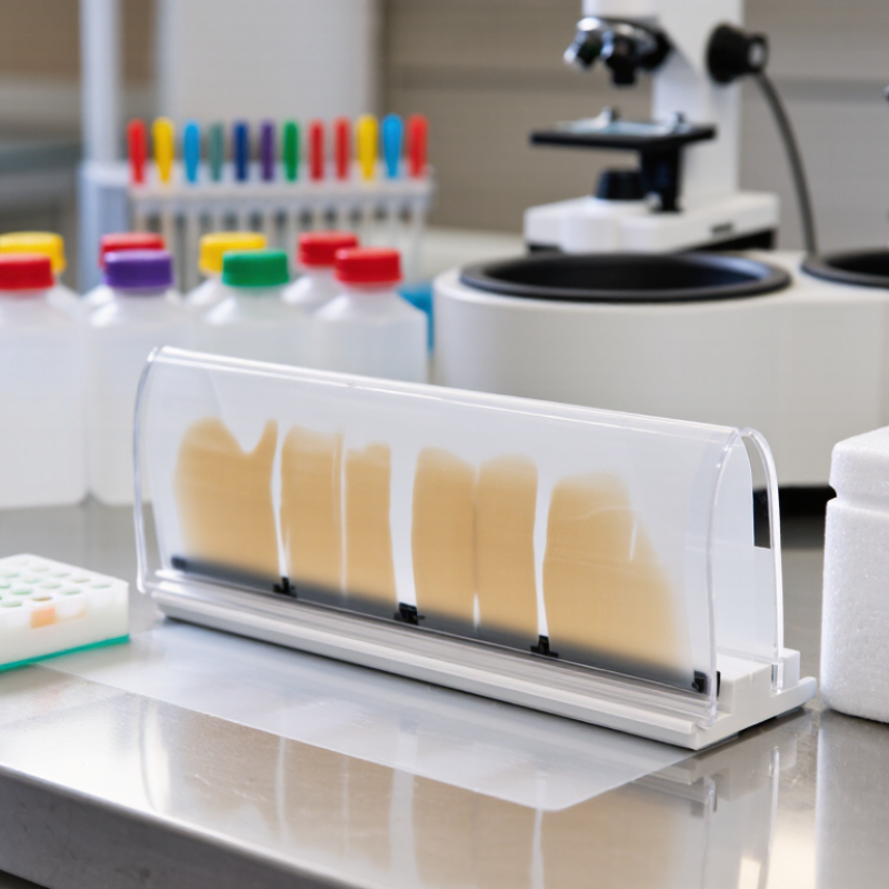
Identify the Antibody’s Target and Intended Experimental Application
Choosing a goat anti-rabbit HRP antibody starts with identifying its real target. It binds rabbit immunoglobulins, not the biological protein studied in your sample. Check the primary antibody’s host species, isotype, and immunoglobulin format. A secondary antibody designed for rabbit IgG may not detect rabbit Fab fragments or unusual isotypes reliably.
The intended application matters just as much. Western blotting uses denatured proteins, while immunohistochemistry and immunofluorescence expose different epitopes inside complex tissue. For ELISA, coating conditions and sample matrices can increase background. Review the datasheet for validated applications, recommended dilution ranges, and cross-reactivity testing. Uhlén and colleagues proposed five validation pillars for antibodies in Nature Methods, including orthogonal and genetic evidence, which supports a stricter selection process. Practical experience also matters: a clean Western blot does not guarantee useful tissue staining.
Control design should match the experiment. Include a secondary-only control, a known positive sample, and an antibody-free negative control. Confirm that endogenous rabbit immunoglobulins are absent or blocked in the specimen. The 2016 Nature reproducibility survey reported that more than 70% of researchers had failed to reproduce another scientist’s experiment. That figure is sobering. I have seen strong HRP signals mistaken for target detection when the real cause was excess secondary antibody. Titrate it carefully, record substrate exposure time, and inspect the membrane before accepting a convincing image. Pretty data can still be wrong.
| Tip | Selection Dimension | What to Verify | Best-Fit Experimental Applications | Practical Selection Guidance |
|---|---|---|---|---|
| 1 | Primary Antibody Species | Confirm that the primary antibody was raised in rabbit. A goat anti-rabbit secondary antibody is designed to recognize rabbit immunoglobulins, including rabbit IgG. | Western blot IHC ICC ELISA | Select goat anti-rabbit HRP only when the primary antibody is rabbit-derived. If the primary antibody is from mouse, rat, or another host, use a species-matched secondary antibody. |
| 2 | Antibody Target and Binding Specificity | Determine whether the reagent recognizes rabbit IgG, the Fc region, or whole immunoglobulin molecules. Check whether cross-reactivity with rabbit IgM, IgA, light chains, or other species has been evaluated. | Routine immunodetection Multiplex assays Low-background workflows | Choose an Fc- or IgG-specific reagent when unwanted binding to other rabbit immunoglobulin classes or immunoglobulin fragments could affect interpretation. |
| 3 | HRP Conjugation and Enzyme Activity | Verify that horseradish peroxidase is directly conjugated to the secondary antibody and that the product has been tested for retained binding and enzymatic activity after conjugation. | Chemiluminescent WB Colorimetric ELISA Chromogenic IHC | Direct HRP conjugation reduces the need for an additional detection step. Select a reagent with validated signal intensity and low nonspecific background in the intended assay. |
| 4 | Experimental Application | Review validation data for the exact technique, including sample type, fixation method, membrane format, blocking conditions, and detection substrate. | Western blot IHC ICC ELISA Dot blot | Do not assume that performance in Western blot will automatically transfer to IHC or ELISA. Prefer application-specific validation whenever possible. |
| 5 | Species Cross-Reactivity | Check the listed reactivity toward rabbit IgG and assess whether the secondary antibody may bind endogenous immunoglobulins in the sample species, such as mouse, human, or bovine proteins. | Animal tissue staining Cell lysates Serum-containing samples | For samples containing immunoglobulins from multiple species, use a highly cross-adsorbed secondary antibody or include appropriate controls to reduce interspecies background. |
| 6 | Conjugate Format and Working Range | Record the recommended dilution range, antibody concentration, formulation, and compatibility with the selected HRP substrate. Common starting dilutions are often around 1:1,000 to 1:10,000 for Western blot, but optimization is required. | Signal optimization Quantitative assays High-sensitivity detection | Start within the validated range and perform a dilution series. Excess secondary antibody can increase background, while insufficient antibody can reduce sensitivity. |
| 7 | Controls, Background, and Storage | Confirm storage temperature, freeze-thaw limits, preservative compatibility, and recommended negative controls. Include a no-primary control and, when appropriate, a rabbit IgG or isotype control. | Reproducible assays Low-background imaging Quantification | Use fresh working dilutions, protect HRP conjugates from contamination and prolonged light exposure, and compare specific signal with control background before drawing conclusions. |
Related Posts
-

China Best Goat Anti Rabbit HRP Suppliers How to Choose?
-
Why Choose PAXgene Blood RNA Tubes for RNA Stability?
-

10 Essential Tips for Using Rabbit Monoclonal Antibodies Effectively?
-
What is a cas9 antibody and how is it used?
-

2026 Best IPSC Cells Innovations for Regenerative Medicine?
-

2026 Top Monoclonal Antibody Innovations and Breakthroughs?